

Researchers at the Laboratory for Photonics and Laser Systems (FOLAS) have studied the generation of cavitation bubbles with a diameter of a few micrometers and for the first time visualized the sharp transition in energy deposition in water by the picosecond laser. The results of the study are published in the journal Ultrasonics Sonochemistry (IF: 9.336).
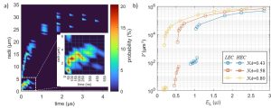
Figure 1: a) Mixed process bubble nucleation and growth observed at laser excitation energy and , b) bubble maximum volume V plotted in regard to excitation laser energy for three focusing numerical apertures. Two groups are marked on the graph. Low energy conversion (LEC) event where only the bubble is detected and high energy conversion (HEC) event where the bubble is detected together with the shock wave and plasma.
When a laser pulse of sufficient energy is focused into water or a water-based medium, a plasma is formed, generally accompanied by a rapid rise in temperature, followed by shock wave emission and cavitation. This converts some of the laser energy into mechanical energy that affects the area outside the laser focal point.
By monitoring the plasma size, cavitation bubble size, and generated shock waves, a twofold threshold process was observed. The identified low energy conversion (LEC) and high energy conversion (HEC) events show a large difference in the energy transferred to the cavitation bubble potential energy with minimal change in excitation laser energy. In the case of LEC events, this leads to reduced mechanical stress on the water-based medium.
The published work adds to the knowledge and research on cavitation caused by laser-induced breakdown (LIB) by studying the bubble dynamics and energy conversion from the excitation source to mechanical energy of a cavitation phenomenon. The results are applicable to laser treatment of biological tissue where highly localized perturbations are desired.
Link to the article: https://doi.org/10.1016/j.ultsonch.2022.106243