

Researchers from the Laboratory for Laser Techniques (LASTEH), in collaboration with the Biotechnical Faculty, Faculty of Medicine, and the Fotona company, studied laser-induced cavitation in wedge-shaped volumes of varying stiffness that simulate conditions in periodontal pockets. The aim of the study was to investigate the possibility of cleaning the infected surfaces of periodontal or peri-implant pockets with laser-induced cavitation. The article was published in the journal Ultrasonics Sonochemistry (IF = 9,336).
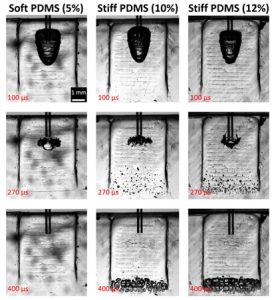
Figure 1: Evolution of cavitation in wedge pockets of different stiffness. On the upper side, a fiber tip can be seen through which laser pulses are introduced, forming the primary cavitation bubble. Due to the pressure shock caused by the collapse of the primary bubble secondary cavitation appears in the bottom part of the pocket. In the case of the soft model, the energy of the primary bubble is dissipated to the deformation of the surrounding tissue, which stops the development of secondary cavitation.
Different laser pulse modalities, PDMS stiffness, and irrigants were tested for their effect on the evolution of cavitation in the narrow wedge geometry. The PDMS stiffness varied in a range that corresponds to severely inflamed, moderately inflamed, or healthy gingival tissue as determined by a panel of dentists. The results imply that deformation of the soft boundary has a major effect on the Er:YAG laser-induced cavitation. The softer the boundary, the less effective the cavitation. We show that in a stiffer gingival tissues model, photoacoustic energy can be guided and focused at the tip of the wedge model, where it enables generation of secondary cavitation and more effective microstreaming. The secondary cavitation was absent in severely inflamed gingival model tissue, but could be induced with a dual-pulse AutoSWEEPS laser modality. This should in principle increase cleaning efficiency in the narrow geometries such as those found in the periodontal and peri-implant pockets and may lead to more predictable treatment outcomes.
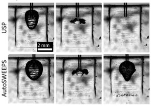
Figure 2: Evolution of cavitation in a soft wedge pocket using single pulse (USP modality) and double pulses (AutoSWEEPS modality).
Link to the article: https://doi.org/10.1016/j.ultsonch.2023.106329.